 3D/4D Image Analysis Software ver10.0
OXFORD IMARIS
Installation date : April 2023(ver10.0 ) October 2019(ver9.3 )
December 2022(ver9.8 ) March 2019(ver9.2 )
November 2019(ver9.5 ) March 2013(ver7.6 )
 Features
 Equipped with the functions necessary to visualize 3D/4D image data obtained with a confocal microscope.
- Capable of creating 3D images from simple rotations to complex animation.
- Animations can be store in QuickTime and AVI format and can be used in presentations.
- The following 2 options are available (added March 2013).
- Measurement Pro: enables various measurements of 3D images such as volume and surface area.
- Track: analyzes changes in objects over time from time-lapse images.
Rules
- You must take a training session.
- Scan digital media (CD, DVD, USB flash drive, etc.) for viruses with the designated computer of our center.
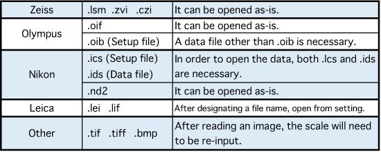
- Zeiss image formats (.lsm, .zvi, and .czi) are recommended but confocal images by other manufacturers can be read as well depending on their formats. Please check the table below.
- Click here for the list of formats available in IMARIS.
-
About training session
- We conduct training sessions as needed. Please contact us in advance to adjust the schedule.
- Please contact us after you have determined when you will start using.
- We only give a rough explanation of how to use the software.
- You will have to analyze your data while receiving advice from the manufacturer. It will take a certain amount of time to master it.
